
Peel our brains with unprecedented precision. Iseult promises, a magnetic resonance imaging (MRI) device designed by the Atomic Energy and Alternative Energy Commission (CEA), whose first anatomical images of the human brain were unveiled on Tuesday 2 April at the Saclay site. In 2021, 11.7 Tesla of its magnetic field has already revealed the inside of the pumpkin, which enters the operational phase after twenty years of development. Since then, CEA teams have cautiously moved to observe their first human subjects.
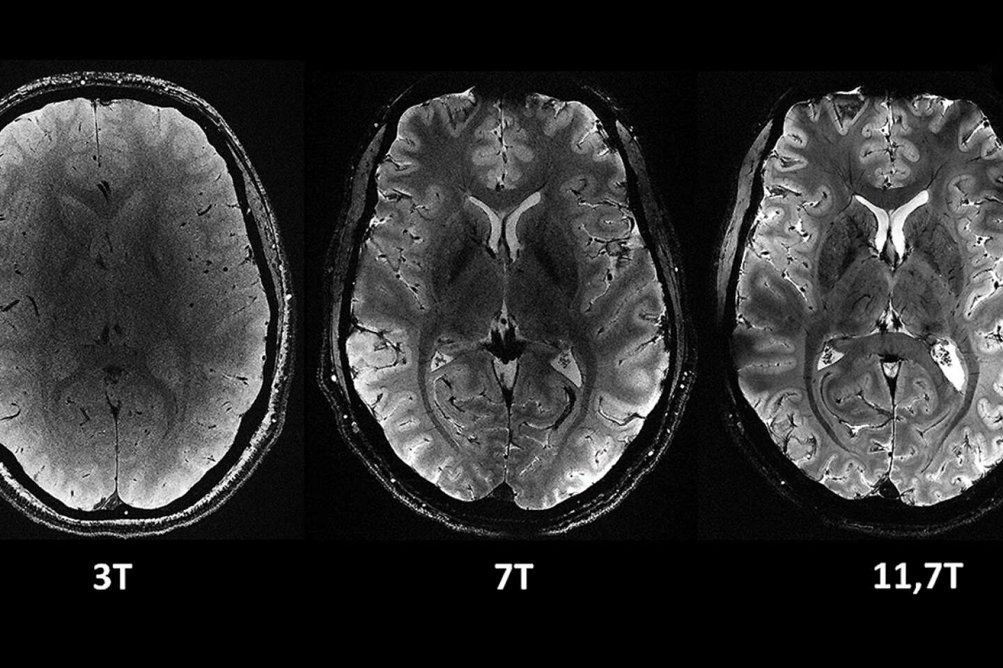
“Images are unbelievably clear! », delighted Nicolas Boulant, responsible for the Iseult project at CEA. A four-minute observation – 0.2 mm in-plane and 1 mm depth – required a two-hour stay in a hospital-quality 3 T scanner – to obtain anatomical images offering the resolution provided by Iseult. A purely theoretical comparison, as patient movement will then blur the image.
If the transition from pumpkin to human took so long, it’s because with Iseult we were entering the uncharted territory of brain discovery. “It was necessary to prove to the health authorities that such an intensity of the magnetic field had no effect on health”, explains Nicholas Boulant. The previous record was 10.5 Tesla on an American machine in Minneapolis. So tests were conducted on 20 healthy adult volunteers to verify Balance, cognition, brain tissue temperature, genotoxicity, etc. A nocébo study was also conducted, in which volunteers were subjected to a session at zero Tesla, without their knowledge, to see if the machine, “that can scare”As Nicholas Boulant notes, psychological bias can be induced. “We saw absolutely nothingAssures the physicist. That was a crucial step to see if we could actually do it A researcher of the human brain. »
“Deciphering the Neural Code”
A new phase will now open, to continue refining the acquisition of data according to the different imaging methods offered by MRI: anatomical data, but also functional data – that is, visualizing brain areas activated by this or that cognitive activity. It will also be possible to carry out so-called “diffusion” imaging, which highlights neuronal bundles connecting different areas of the brain. The intensity of the magnetic field should also make it possible to detect compounds invisible at lower fields, such as lithium, used in bipolar disorders, and glucose and glutamate, small molecules involved in brain metabolism.
You have 44.61% of this article left to read. The rest is reserved for subscribers.